
Peter Ciesielski
Researcher V-Materials Science
Peter.Ciesielski@nrel.gov
303-384-7691
 https://orcid.org/0000-0003-3360-9210
Google Scholar
Research Interests
I am a scientist at the National Renewable Energy Laboratory (NREL) and have an interdisciplinary background and training. My research encompasses many aspects of bioenergy and biomaterials science. Biomass is an abundant, renewable resource that is naturally and continually mass-produced on this planet. Humans have been using biomass as source of fuel and materials for as long as they have existed. My research aims to improve ways by which biomass can be used as a sustainable and renewable source of fuels, chemicals, and materials.
Microscopy and Structural Modeling
Microscopy is an important and powerful tool for investigating the spatial distribution of mass. While microscopy has often been considered to provide predominantly qualitative information, microscopy data may be subject to rigorous quantitative analysis, as would any other experimental technique. Shown below are examples of images of various samples at scales of different length using several different imaging techniques.
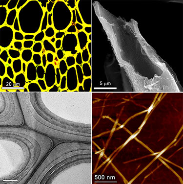
Examples of multi-modal imaging of plant cell walls. (Top left) Confocal scanning laser microscopy of cell walls in cross-section. (Top right) Scanning electron microscopy of a broken fiber cell. (Bottom left) Transmission electron microscopy of several secondary cell walls. (Bottom right) Atomic force microscopy of algal cellulose nanofibrils.
Transmission Electron Tomography and Nanoscale/Atomic Modeling
Perhaps the most powerful tool for nanostructural characterization is 3-D electron tomography. I learned this from technique from Bryon Donohoe, who is a great microscopist at NREL. Recently, we used this technique to visualize and model the nanostructure of thermochemically treated biomass. Below is one of the tomograms that resulted from this study, visualized by panning through the volume in the z dimension. The scale bar is 200 nm. (This is one of my favorite tomograms because it really shows the fibrous structure of the cellulose in the wall.)
Tomographic volume of steam-exploded corn stover cell wall. This movie visualizes a volume reconstruction of steam-exploded corn stover cell walls by panning through the z-dimension of the volume.
I developed some novel quantitative analysis methods, implemented in MATLAB, to discern the longitudinal geometry of cellulose microfibrils within the tomographic data and used the results to construct and evaluate atomic models for the cellulose microfibrils (Ciesielski et al., ACS Nano, 2013). A movie visualizing how these macromolecular models are built from the unit cell of a 36-chain cellulose fibril is shown below.
Construction of atomic models of cellulose nanofibrils along an arbitrary space curve. This movie visualizes how the unit cell of a cellulose crystal may be used to build atomic models of cellulose fibrils with an arbitrary longitudinal axis. In this model, the Frenet-Seret frame of the space curve is used to orient the unit cell to align the 100-crystallographic direction with the unit normal vector.
Examples of atomic models of cellulose constructed in this manner are shown below. (Atomic models are shown as blue and black spheres; the measured tomographic density is shown as the semi-transparent light brown isosurface.)
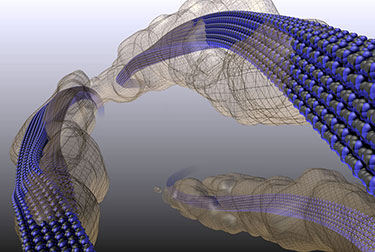
Atomic models of cellulose nanofibrils informed by TEM tomography.
Micro/Mesoscale Structural Modeling
We are also developing methods to model the higher-order architecture of biomass and plant cell walls. I use a few different tools to do this, all of which employ Constructive Solid Geometry (CSG) in some way. Plant cell walls provide an interesting example of mesostructure, wherein the assembly of cellulose nanofibrils and other biopolymers form a robust composite with excellent mechanical properties.
Below is an image of one of the first 3-D cell wall mesostructure models I constructed. This was built and visualized with POVRay. This model was basically just for visualization but still employed a CSG algorithm to produce the model based on quantitative parameters that described the composition and geometry of the components.
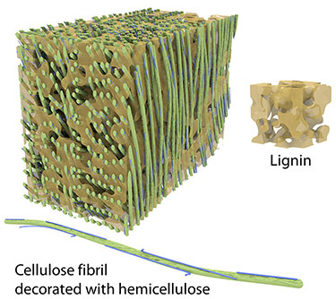
3-D model of cell wall mesostructure. This is a visualization of a rectangular chunk of a lignocellulosic cell wall. All the parameters that dictate the structure of the model, such as nanofribril spacing, curvature, hemicellulose decoration, lignin porosity, etc., are variables in the model construction algorithm.
Recently we have extended the CSG algorithm to produce density field models of lignocellulose that can be evaluated with finite element simulations. Currently we are performing simulations of structural mechanics and transport phenomena with these models to investigate how variations in the structure and composition of lignocellulose impacts its functionality in scenarios relevant to biomass conversion processes. Below is a movie that visualizes a density field model for lignocellulose mesostructure.
The microstructure of biomass is also an important feature that has a big impact on bulk properties and that can vary substantially among plant species. We use multimodal imaging and quantitative image analysis to measure geometric parameters that specify the microstructure of biomass, including cell wall thickness, cell lumen diameter, and spatial arrangement and frequency of various cell types. I wrote a CSG algorithm to take these parameters, along with others that describe the size and shape of the whole particle, and produced a 3-D model. These methods were published recently (Ciesielski, et. al., Energy & Fuels, 2015). These were the first biomass particle models to incorporate explicit, species-specific microstructure. Examples of these models are shown in the movie below.
Biomass particle models of three different sizes. This video visualizes biomass particle models constructed from structural data obtained from a milled poplar feedstock. The sizes and shapes of these models are representative of populations of particles within the feedstock ensemble.
Finite Element Simulation of Transport Phenomena and Biomass Conversion Processes
Biomass Particle Modeling
Biomass particles used as feedstocks for both thermochemical and biochemical conversion processes retain the microstructural anatomy of the plant of origin, which varies substantially among species. Furthermore, when biomass is milled prior to conversion, a distribution of particle sizes is produced. Both of these factors can have a significant impact on intra-particle transport phenomena (i.e., transfer of heat, mass, and momentum), which are critical processes that affect the observed rates of catalytic conversion processes.
The vast majority of biomass particle models present in the literature make many simplifying assumptions about the geometry of the particles. While these "low order" models are sometimes sufficient to fit data from a specific experiment, reactor, or feedstock, they are of little value for predicting the behavior of different feedstocks or feedstock blends in arbitratry reactors and operating conditions. We are developing methods to produce more realistic, species-specific particle models that predict how a given biomass feedstock or feedstock blend will perform in various conversion processes. Examples of these models are shown in the figure below, alongside SEM micrographs of real biomass particles that the models are intended to represent. These models are used in multiphysics, finite element simulations of transport phenomena coupled to chemical conversion kinetics to investigate the behavior of these particles in various simulations. An example of a meshed particle model is shown in the middle row of the figure below. Since this effort focuses on modeling the biomass rather than a specific reactor or process, these structural models are relevant to any process that takes biomass as a feedstock. The bottom row of the figure below shows example snapshots from simulations of conjugate heat transfer, showing slower heating behavior of a hardwood particle compared to a softwood particle of the same size due to differences in microstructure. All of the images in this figure were adapted from Ciesielski, et al., Energy & Fuels, 2015.
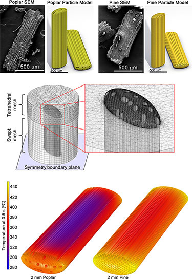
Finite element modeling of transport phenomena in biomass particles with realistic morphology and microstructure. (Top) Comparison of 3-D biomass particle models to scanning electron micrographs of the same. (Middle) Finite element mesh of a complex particle model. (Bottom) Temperature profiles predicted by simulations of conjugate heat transfer.
Catalyst Particle Modeling
I am also starting to apply similar techniques to model transport phenomena and reaction kinetics within structured catalysts. The figure below illustrates how a transmission electron micrograph of a zeolite catalyst particle, which is an agglomerate of many small zeolite crystallites, may be used to generate geometry for a finite element simulation.
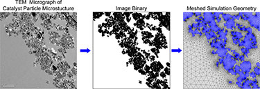
Importing microscopy data of Zeolite particle microstructure to serve as the geometry for finite element simulation. (Left) TEM micrograph of the edge of catalyst particle, showing a cluster of small crystallites. (Middle) A binary image obtained from the TEM micrograph. (Right) Meshed simulation geometry.
These geometries are used to simulate transport and chemical transformations of reactants and products during catalytic upgrading. The movie below shows the localized product formation predicted by such a simulation. Notice how the product concentration builds inside the catalyst particle due to mass transport limitations.
Product formation within a microstructured Zeolite catalyst particle.
Molecular Dynamics Simulation
This subject has been a long-time research interest of mine, which is to say that I have been very interested in molecular dynamics simulations performed by other researchers. I have recently begun to write some of my own MD simulations, with much help and guidance from Michael Crowley, who is a CHARMM developer and fantastic scientist here at NREL. Below is a movie from one of my first MD simulations, showing the relaxation of a curved cellulose nanofibril. This simulation was written in CHARMM and visualized with Pymol.
Molecular dynamics simulation of a cellulose nanofibril relaxing from a strained configuration.
Production and Applications of Nanocellulose and Glycomaterials
Nanocellulose is an impressive class of renewable materials with outstanding properties and incredible potential in many applications including advanced polymer composites, films and foams, biomedical and biocompatible materials, and green electronics and photonics. These materials are derivatives of cellulose naturally synthesized by plants (though cellulosic nanomaterials can also be produced from bacterial and algal sources). Because the energy to produce these materials comes from sunlight and the carbon comes from CO2 (in the case of plant and algal sources), they are an excellent energy-efficient and sustainable alternative to many non-renewable and/or environmentally detrimental petroleum-derived materials. We are investigating ways to co-produce nanocellulose and biofuels in a format compatible with lignocellulosic (2nd generation) biorefinery technology. We are also pursuing a variety of end-use applications for nanocellulose and other plant-derived glycopolymeric materials.
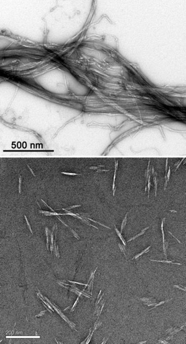
TEM images of cellulose nanofibrils (CNFs, top) and cellulose nanocrystals (CNCs, bottom) produced by the USDA Forest Products Lab and imaged in the NREL Biomass Surface Characterization Laboratory.
Scientific Visualization
I spend a lot of time generating figures and schematics to visualize scientific data and concepts, mostly for publications in journals and reports. High-quality visualization is both an art and a science. I don't have any official training in this area, but I have learned to use some pretty nice visualization software like POVRay, Blender, and Pymol. I've included a few of my favorite images here. One of the best things about a lot of the structures and models visualized here is that they are more than just pictures; they are actually functional models that can be evaluated in computational environments.
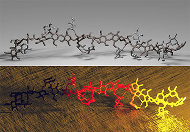
Hemicellulose Molecular Models. These are molecular models built in CHARMM by Michael Crowley at NREL, rendered by Peter Ciesielski in POVRay. The top image uses several different stone textures, and the bottom image uses emission media to make the molecule appear as if it just came out of a forge and is starting to cool.
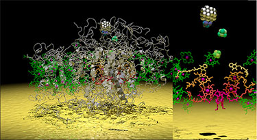
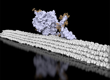
Platinized Photosystem I on a Gold Surface. This is a visualization of the molecular structure of Photosystem I (one of the incredibly important proteins that participates in the light reactions of photosynthesis), which has a platinum nanocluster sitting on its terminal electron donor site. Some artistic license was taken for the platinum cluster, but the atomic structure of PSI was obtained by X-ray crystallography by Amunts, et al. The right panel shows a zoom view of the electron transfer chain with the protein scaffolding hidden. What an awesome nanomachine! Some version of this figure with additional annotation will appear in a book chapter soon. I will link to it when it's available. This figure was generated using Pymol and POVRay.
Cel7A (or CBH1, if you prefer) on cellulose fibril. This image was created from a snapshot of a molecular dynamics simulation written by Gregg Beckham to investigate interactions between this cellulase enzyme and its cellulose substrate. The figure was generated using Pymol and POVRay.
Affiliated Research Programs
-
Microscopy and Structural Modeling
-
Finite Element Simulation of Transport Phenomena and Biomass Conversion Processes
-
Molecular Dynamics Simulation
-
Production and Applications of Cellulose Nanocrystals
-
Scientific Visualization
Areas of Expertise
-
Advanced Multimodal Microscopy: Scanning and transmission electron microscopies; 3-D electron tomography; atomic force microscopy; confocal Raman, florescence, and energy dispersive X-Ray hyper-spectral imaging.
-
Quantitative Image Analysis and Structural Modeling: Development and implementation of computational methods for geometric analysis of tomographic reconstructions; semi-automated image analysis and computer vision; atomistic modeling of plant biopolymers; micro- and nonstructural modeling of plant cell walls, biomass particles, biopolymers, and inorganic materials.
-
Surface Science and Electrochemistry: Inorganic surface modification and characterization; analytical electrochemistry and electrochemical catalysis; surface functionalization for biological compatibility and protein adsorption; fabrication of biohybrid systems and devices.
-
Computational Modeling, Simulation, and Visualization: Kinetic modeling of catalytic and photo-catalytic reactions (MATLAB, Python); finite element simulation of transport phenomena in complex geometries (COMSOL); molecular dynamics simulation (CHARMM); computational visualization and ray-tracing (POVRay, Blender).
Education
Ph.D., Interdisciplinary Material Science, Vanderbilt University, 2010
B.S., Chemical and Biological Engineering, Colorado State University, 2006
Featured Work
"Multiscale Alterations in Sugar Cane Bagasse and Straw Submitted to Alkaline Deacetylation," ACS Sustainable Chemistry and Engineering (2018)
"Enhanced mobility CsPbI3 quantum dot arrays for record-efficiency, high-voltage photovoltaic cells," Science Advances (2017)
"Lignocellulose deconstruction in the biosphere," Current Opinion in Chemical Biology (2017)
"Estimating the Temperature Experienced by Biomass Particles during Fast Pyrolysis Using Microscopic Analysis of Biochars," Energy & Fuels (2017)
"Understanding Trends in Autoignition of Biofuels: Homologous Series of Oxygenated C5 Molecules," The Journal of Physical Chemistry A (2017)
"Multifunctional Cellulolytic Enzymes Outperform Processive Fungal Cellulases for Coproduction of Nanocellulose and Biofuels," ACS Nano (2017)
"Catalyst Residence Time Distributions in Riser Reactors for Catalytic Fast Pyrolysis. Part 2: Pilot-Scale Simulations and Operational Parameter Study," ACS Sustainable Chemistry and Engineering (2017)
"Predictive Model for Particle Residence Time Distributions in Riser Reactors. Part 1: Model Development and Validation," ACS Sustainable Chemistry and Engineering (2017)
"Estimation of Heat Transfer Coefficients for Biomass Particles by Direct Numerical Simulation Using Microstructured Particle Models in the Laminar Regime," ACS Sustainable Chemistry & Engineering (2017)
"Directed plant cell-wall accumulation of iron: embedding co-catalyst for efficient biomass conversion," Biotechnology for Biofuels (2016)
"Not Just Lumber—Using Wood in the Sustainable Future of Materials, Chemicals, and Fuels," Journal of Materials (2016)
"Influence of Crystal Allomorph and Crystallinity on the Products and Behavior of Cellulose during Fast Pyrolysis," ACS Sustainable Chemistry & Engineering (2016)
"Wood-derived materials for green electronics, biological devices, and energy applications," Chemical Reviews (2016)
"Development of Lignocellulosic Biorefinery Technologies: Recent Advances and Current Challenges," Australian Journal of Chemistry (2016)
"Low-Order Modeling of Internal Heat Transfer in Biomass Particle Pyrolysis," Energy & Fuels (2016)
"cis, cis-Muconic acid: separation and catalysis to bio-adipic acid for nylon-6, 6 polymerization," Green Chemistry (2016)
"Pilot-Scale Batch Alkaline Pretreatment of Corn Stover," ACS Sustainable Chemistry & Engineering (2015)
"Biomass accessibility analysis using electron tomography," Biotechnology for Biofuels (2015)
"Improving Sugar Yields and Reducing Enzyme Loadings in the Deacetylation and Mechanical Refining (DMR) Process through Multistage Disk and Szego Refining and Corresponding Techno-Economic Analysis," ACS Sustainable Chemistry & Engineering (2015)
"Manipulation of guaiacyl and syringyl monomer biosynthesis in an Arabidopsis cinnamyl alcohol dehydrogenase mutant results in atypical lignin biosynthesis and modified cell wall structure," The Plant Cell (2015)
"Alkaline pretreatment of switchgrass," ACS Sustainable Chemistry & Engineering (2015)
"Identifying the ionically bound cell wall and intracellular glycoside hydrolases in late growth stage Arabidopsis stems: implications for the genetic engineering of bioenergy crops," Frontiers in Plant Science (2015)
"Transgenic ferritin overproduction enhances thermochemical pretreatments in Arabidopsis," Biomass and Bioenergy (2015)
"Molybdenum incorporated mesoporous silica catalyst for production of biofuels and value-added chemicals via catalytic fast pyrolysis," Green Chemistry (2015)
"Biomass Particle Models with Realistic Morphology and Resolved Microstructure for Simulations of Intraparticle Transport Phenomena," Energy & Fuels (2015)
"Metal-silicate catalysts: Single site, mesoporous systems without templates," J. Nanosci. Lett. (2015)
"Disruption of Mediator rescues the stunted growth of a lignin-deficient Arabidopsis mutant," Nature (2014)
"The Structure of the catalytic domain of a plant cellulose synthase and its assembly into dimers," The Plant Cell Online (2014)
"Engineering plant cell walls: tuning lignin monomer composition for deconstructable biofuel feedstocks or resilient biomaterials," Green Chemistry (2014)
"Alkaline pretreatment of corn stover: Bench-scale fractionation and stream characterization," ACS Sustainable Chemistry & Engineering (2014)
"Effect of mechanical disruption on the effectiveness of three reactors used for dilute acid pretreatment of corn stover Part 2: morphological and structural substrate analysis," Biotechnol. Biofuels (2014)
"Effect of mechanical disruption on the effectiveness of three reactors used for dilute acid pretreatment of corn stover Part 1: chemical and physical substrate analysis," Biotechnol. Biofuels (2014)
"Clean fractionation pretreatment reduces enzyme loadings for biomass saccharification and reveals the mechanism of free and cellulosomal enzyme synergy," ACS Sustainable Chemistry & Engineering (2014)
"Hydrothermal catalytic processing of saturated and unsaturated fatty acids to hydrocarbons with glycerol for in situ hydrogen production," Green Chemistry (2014)
"Evaluation of clean fractionation pretreatment for the production of renewable fuels and chemicals from corn stover," ACS Sustainable Chemistry & Engineering (2014)
"Multiscale deconstruction of molecular architecture in corn stover," Scientific Reports (2014)
"Lignin depolymerisation by nickel supported layered-double hydroxide catalysts," Green Chemistry (2014)
"Real-time monitoring of the deactivation of HZSM-5 during upgrading of pine pyrolysis vapors," Green Chemistry (2014)
"Chemically induced conditional rescue of the reduced epidermal fluorescence8 mutant of Arabidopsis reveals rapid restoration of growth and selective turnover of secondary metabolite pools," Plant Physiology (2014)
"3D electron tomography of pretreated biomass informs atomic modeling of cellulose microfibrils," ACS Nano (2013)
"Mechanistic study of a ru-xantphos catalyst for tandem alcohol dehydrogenation and reductive aryl-ether cleavage," ACS Catalysis (2013)
"Elucidating the role of ferrous ion cocatalyst in enhancing dilute acid pretreatment of lignocellulosic biomass," Biotechnol. Biofuels (2011)
"Kinetic model of the photocatalytic effect of a photosystem I monolayer on a planar electrode surface," J. Phys. Chem. A. (2011)
"Patterned nanoporous gold as an effective SERS template," Nanotechnology (2011)
"Enhanced photocurrent production by photosystem I multilayer assemblies," Advanced Functional Materials (2010)
"Photosystem I-based biohybrid photoelectrochemical cells," Bioresource Technology (2010)
"Leaving their legacy: A research experience and teaching module to maximize STEM learning for students," The Science Teacher (2010)
"Functionalized nanoporous gold leaf electrode films for the immobilization of photosystem I," ACS Nano (2008)
"Rapid assembly of photosystem I monolayers on gold electrodes," Langmuir (2008)
Book Chapters
"Simulating Biomass Fast Pyrolysis at the Single Particle Scale," in Fast Pyrolysis of Biomass (2017)
"Photobiohybrid Solar Conversion with Metalloenzymes and Photosynthetic Reaction Centers," in Biotechnologies for Biofuel Production and Optimization (2016)
"Preservation and Preparation of Lignocellulosic Biomass Samples for Multi-scale Micropscopy and Analysis," Biomass Conversion: Methods and Protocols (2012)
The images, graphics, and movies on this page were generated by Peter Ciesielski at NREL. If you wish to use these images, please contact Peter directly.
Share
