Scanning Electron Microscopy Basic Operating Mode
NREL uses scanning electron microscopy in its basic operating mode to sense secondary electrons and X-rays emitted from a materials sample.
The former provides high-resolution topography, and the latter indicates elemental composition.
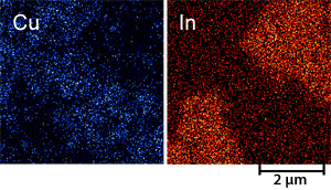
CuInSe2 thin film. Cu and In elemental maps obtained by EDS secondary electrons, with energies in the range of 0 to 50 eV, provide an excellent topographic image of the surface under observation with very high resolution and depth of field at very high scanning speeds.
Backscattered electron emission is sensitive to composition. Energy transfer from incident electrons to the specimen produces X-rays characteristic of the specimen's elemental composition by energy-dispersive spectroscopy (EDS). Elements are identified on the energy-resolved spectrum of X-rays by their characteristic lines. Synchronizing the scanning of the electron beam with the readout of the EDS detector can generate elemental maps with micrometric resolution. Under well-controlled conditions, the composition of the specimen can be quantified with an accuracy of 1%–3%.
Share